Introduction to Posterior Vitreous Detachment
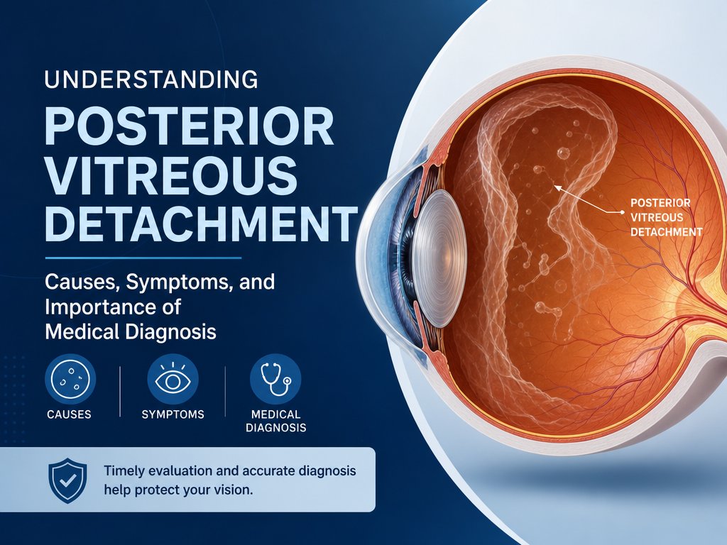
Posterior vitreous detachment (PVD) is a prevalent eye condition associated with aging. It involves the vitreous, a gel-like substance in the eye, shrinking and detaching from the retina. Although this condition is a natural part of aging and generally benign, it can sometimes lead to complications. Understanding the condition is essential, as it helps in recognizing changes in vision and knowing when to seek medical advice.
What Happens in PVD?
The vitreous is a gel that fills the space between the lens and the retina. When PVD occurs, this gel contracts and pulls away from the retina. This process itself does not usually result in vision loss, and many individuals experience no harmful effects. However, in rare cases, it can lead to retinal detachment, which requires medical intervention. This potential risk underscores the importance of monitoring symptoms carefully.
Causes and Risk Factors
Aging is the primary factor causing PVD. The vitreous loses its consistency, making detachment more likely as one grows older, typically after 60. Although less common, PVD can occur in individuals younger than 40, especially in those with certain risk factors, including eye surgery, significant eye trauma, or conditions like myopia. Genetic predispositions and certain health conditions may also influence the likelihood of developing PVD. People with diabetes or cataracts are sometimes at increased risk.
Recognizing Symptoms
PVD itself is painless and not life-threatening. However, symptoms like new floaters, flashes of light, or a shadow appearing in the vision field may arise. These are signals indicating potential issues warranting a medical check-up. Such symptoms might herald either PVD or more serious conditions such as retinal detachment, vitreous hemorrhage, or inflammation, each requiring a different approach to treatment.
Diagnosis
Diagnosis of PVD involves an eye examination where the doctor dilates the pupils to inspect the retina and optic nerve thoroughly. Procedures like optical coherence tomography or ocular ultrasound might be employed for a clearer view if the vitreous is very transparent or if the detachment is not easily visible during the initial examination. These tools provide detailed images necessary for assessing the degree of PVD and any possible threats to the retina.
Treatment Considerations
In most cases, no treatment is necessary for PVD. The condition often resolves without intervention, as the eye adjusts to the changes naturally over time. However, if symptoms persist or new vision problems develop even after detachment is complete, further medical evaluation may be required. Seek immediate medical attention for sudden vision changes, an increase in floaters, or flashes, as these may indicate more serious issues like retinal tears or detachment, which can impair vision if not treated.
Importance of Regular Eye Examinations
Regular eye exams are crucial for early detection and management of PVD and other ocular conditions. They provide an opportunity for eye care professionals to monitor eye health over time and identify changes that might require action. Avoid self-diagnosing and consult an eye healthcare professional for accurate diagnosis and tailored treatment plans, ensuring the best care and prevention of possible complications. Also, regular exams are beneficial in monitoring overall eye health and detecting other issues that might arise.
In summary, while PVD is a common condition associated with aging and usually not serious, understanding its symptoms and seeking timely medical advice can prevent potential complications. Awareness and education on eye health contribute significantly to maintaining quality vision over one’s lifetime. Prioritizing eye health can have a profound effect on maintaining independence and quality of life as people age.